
پرونده:Hepatocellular carcinoma 1.jpg
Hepatocellular_carcinoma_1.jpg (۵۵۰ × ۳۶۸ پیکسل، اندازهٔ پرونده: ۳۸ کیلوبایت، نوع MIME پرونده: image/jpeg)
این پرونده در ویکیانبار موجود است. محتویات صفحهٔ توصیف آن در زیر نمایش داده میشود. |
خلاصه
| توضیح |
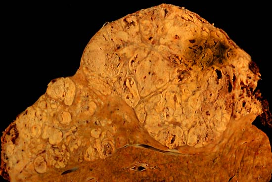
Hepatocellular carcinoma This specimen is from a 50ish woman who presented to the hospital with abdominal pain and ascites. The radiologist recovered what appeared to be whole blood on paracentesis. Cytological exam of the bloody fluid showed no evidence of malignancy. Liver function tests were abnormal, and serologic tests were positive for antibody to hepatitis C. The patient deteriorated rapidly and died within a few days. The autopsy showed this hepatocellular carcinoma occupying much of the volume of a cirrhotic liver. Furthermore, the tumor had invaded the diaphragm and ruptured into the peritoneal cavity, causing the bloody ascites. The photo shows a view of a longitudinal slice taken through the full length of the liver. The photos were shot with a Minolta X-370 with 100 mm bellows lens on Kodak Elite ISO 100 transparency film. The specimen was sliced fresh and fixed in formalin overnight, then briefly immersed in 70% alcohol to retrieve some of the native color and dull the surface reflections. Photograph by Ed Uthman, MD. Public domain. Posted 23 Sep 00 |
| منبع | http://web2.airmail.net/uthman/specimens/index.html |
| پدیدآور | |
| اجازهنامه (استفادهٔ مجدد از این پرونده) |
PD |
اجازهنامه
| این اثر توسط پدیدآور آن، Ed Uthman، به مالکیت عمومی درآمده است. این مربوط به تمام جهان است. در برخی از کشورها ممکن است به صورت قانونی این امکانپذیر نباشد؛ اگر چنین است: Ed Uthman به هر کسی اجازهٔ استفاده از این اثر برای هر مقصودی، بدون هیچگونه شرایطی، را میدهد تا وقتی که این شرایط توسط قانون مستلزم نشده باشند.
|
تاریخچهٔ پرونده
روی تاریخ/زمانها کلیک کنید تا نسخهٔ مربوط به آن هنگام را ببینید.
| تاریخ/زمان | بندانگشتی | ابعاد | کاربر | توضیح | |
|---|---|---|---|---|---|
| کنونی | ۵ ژوئن ۲۰۰۶، ساعت ۱۰:۱۴ | | ۵۵۰ در ۳۶۸ (۳۸ کیلوبایت) | Patho | {{Information| |Description=Hepatocellular carcinoma This specimen is from a 50ish woman who presented to the hospital with abdominal pain and ascites. The radiologist recovered what appeared to be whole blood on paracentesis. Cytological exam of the blo |
کاربرد پرونده
صفحههای زیر از این تصویر استفاده میکنند:
کاربرد سراسری پرونده
ویکیهای دیگر زیر از این پرونده استفاده میکنند:
- کاربرد در ar.wikipedia.org
- کاربرد در ast.wikipedia.org
- کاربرد در az.wikipedia.org
- کاربرد در be.wikipedia.org
- کاربرد در bs.wikipedia.org
- کاربرد در ca.wikipedia.org
- کاربرد در cs.wikipedia.org
- کاربرد در de.wikipedia.org
- کاربرد در de.wikibooks.org
- کاربرد در el.wikipedia.org
- کاربرد در en.wikipedia.org
- Hepatocellular carcinoma
- Alcohol and cancer
- Portal:Medicine/Selected article/50, 2007
- Portal:Medicine/Selected Article Archive (2007)
- Obesity-associated morbidity
- Cirrhosis
- Portal:Viruses
- Portal:Viruses/Selected article
- Portal:Viruses/Selected article/10
- User:Daniel Mietchen/Wikidata lists/Items with Disease Ontology IDs
- کاربرد در eo.wikipedia.org
- کاربرد در es.wikipedia.org
- کاربرد در eu.wikipedia.org
- کاربرد در fi.wikipedia.org
- کاربرد در fr.wikipedia.org
- کاربرد در gl.wikipedia.org
- کاربرد در he.wikipedia.org
- کاربرد در hi.wikipedia.org
- کاربرد در hy.wikipedia.org
- کاربرد در id.wikipedia.org
- کاربرد در it.wikipedia.org
- کاربرد در ja.wikipedia.org
- کاربرد در kk.wikipedia.org
نمایش استفادههای سراسری از این پرونده.