پرش به محتوا

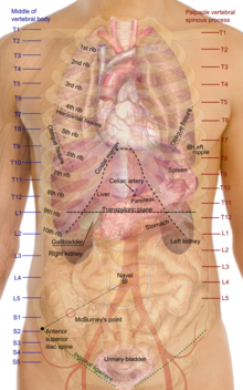
پرونده:Surface projections of the organs of the trunk.svg

محتوای صفحه در زبان‌های دیگر پشتیبانی نمی‌شود
از ویکی‌پدیا، دانشنامهٔ آزاد

پروندهٔ اصلی(پروندهٔ اس‌وی‌جی، با ابعاد ۴۷۵ × ۷۶۰ پیکسل، اندازهٔ پرونده: ۳٫۲ مگابایت)

خلاصه

توضیح
English: Surface projections of the major organs of the trunk, using the vertebral column and rib cage as main reference sources found in Superficial anatomy. The transpyloric plane and McBurney's point are among the marked locations.

For reference list and more information, see list on main page for the png-version

To discuss image, please see Talk:Human body diagrams

تاریخ
منبع All used images are in public domain.
پدیدآور Mikael Häggström
دیگر نسخه‌ها
.png-format.
Without labels.

اجازه‌نامه

Public domain من، دارنده حق تکثیر این اثر، این اثر را به مالکیت عمومی منتشر می‌کنم. این قابل اجرا در تمام نقاط جهان است.
در برخی از کشورها ممکن است به صورت قانونی این امکان‌پذیر نباشد؛ اگر چنین است:
من اجازهٔ استفاده از این اثر را برای هر مقصودی، بدون هیچ‌گونه شرایطی می‌دهم، تا وقتی که این شرایط توسط قانون مستلزم نشده باشد.

Human body diagrams

Main article at: Human body diagrams

Template location:Template:Human body diagrams

How to derive an image

Derive directly from raster image with organs

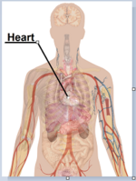
The raster (.png format) images below have most commonly used organs already included, and text and lines can be added in almost any graphics editor. This is the easiest method, but does not leave any room for customizing what organs are shown.

Adding text and lines:

Derive "from scratch"

By this method, body diagrams can be derived by pasting organs into one of the "plain" body images shown below. This method requires a graphics editor that can handle transparent images, in order to avoid white squares around the organs when pasting onto the body image. Pictures of organs are found on the project's main page. These were originally adapted to fit the male shadow/silhouette.

Organs:

More organs are found at: Human body diagrams/Organs

Derive by vector template

The Vector templates below can be used to derive images with, for example, Inkscape. This is the method with the greatest potential.

See Human body diagrams/Inkscape tutorial for a basic description in how to do this.

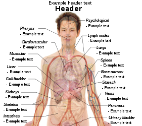
Examples of derived works

More examples

Licensing

Creative Commons CC-Zero این پرونده تحت CC0 1.0 Universal Public Domain Dedication کریتیو کامنز قابل دسترسی است.
کسی که اثری را با این سند همراه کرده است، با چشم‌پوشی از تمام حقوق خود نسبت به اثر در جهان تحت قانون کپی‌رایت و همهٔ حقوق قانونی مرتبط یا همسایه‌ای که او در اثر داشته است، تا حد مجاز در قانون، آن را به مالکیت عمومی اهدا کرده است. شما می‌توانید بدون گرفتن اجازه این اثر را تکثیر کنید، تغییر دهید، منتشر کنید یا دوباره ایجاد کنید، حتی اگر مقاصد تجاری داشته باشید.

عنوان

شرحی یک‌خطی از محتوای این فایل اضافه کنید

آیتم‌هایی که در این پرونده نمایش داده شده‌اند

توصیف‌ها

تاریخچهٔ پرونده

روی تاریخ/زمان‌ها کلیک کنید تا نسخهٔ مربوط به آن هنگام را ببینید.

(جدیدترین | قدیمی‌ترین) نمایش (۱۰ مورد جدیدتر  | ) (۱۰ | ۲۰ | ۵۰ | ۱۰۰ | ۲۵۰ | ۵۰۰)
تاریخ/زمانبندانگشتیابعادکاربرتوضیح
کنونی‏۲۷ دسامبر ۲۰۱۹، ساعت ۰۹:۱۹تصویر بندانگشتی از نسخهٔ مورخ ‏۲۷ دسامبر ۲۰۱۹، ساعت ۰۹:۱۹۴۷۵ در ۷۶۰ (۳٫۲ مگابایت)Mikael Häggström+Costal margin
‏۱۱ نوامبر ۲۰۱۰، ساعت ۱۰:۳۸تصویر بندانگشتی از نسخهٔ مورخ ‏۱۱ نوامبر ۲۰۱۰، ساعت ۱۰:۳۸۴۷۵ در ۷۶۰ (۳٫۲ مگابایت)Mikael HäggströmAdapted to recently added overview images. Distinguished different ways to designate vertebrae levels.
‏۷ نوامبر ۲۰۱۰، ساعت ۱۰:۰۴تصویر بندانگشتی از نسخهٔ مورخ ‏۷ نوامبر ۲۰۱۰، ساعت ۱۰:۰۴۴۶۰ در ۷۴۰ (۳٫۱۶ مگابایت)Mikael Häggströmheart in from of liver
‏۷ نوامبر ۲۰۱۰، ساعت ۰۹:۴۱تصویر بندانگشتی از نسخهٔ مورخ ‏۷ نوامبر ۲۰۱۰، ساعت ۰۹:۴۱۴۶۰ در ۷۴۰ (۳٫۱۶ مگابایت)Mikael HäggströmMade heart a bit smaller. Marked sacral levels.
‏۴ نوامبر ۲۰۱۰، ساعت ۱۶:۵۷تصویر بندانگشتی از نسخهٔ مورخ ‏۴ نوامبر ۲۰۱۰، ساعت ۱۶:۵۷۴۶۰ در ۷۴۰ (۳٫۰۹ مگابایت)Mikael HäggströmLowered urinary bladder according to Fig 139 (see ref list)
‏۲۶ اکتبر ۲۰۱۰، ساعت ۰۴:۲۸تصویر بندانگشتی از نسخهٔ مورخ ‏۲۶ اکتبر ۲۰۱۰، ساعت ۰۴:۲۸۴۵۰ در ۷۳۲ (۳٫۰۴ مگابایت)Mikael Häggströmsymmetry
‏۲۶ اکتبر ۲۰۱۰، ساعت ۰۴:۲۶تصویر بندانگشتی از نسخهٔ مورخ ‏۲۶ اکتبر ۲۰۱۰، ساعت ۰۴:۲۶۴۵۰ در ۷۳۲ (۳٫۰۴ مگابایت)Mikael HäggströmMoved T and L labels more to the right. Raised vertebral ending of some ribs to reach their correct level. Raised "stomach" label to avoid contact with the T10 line.
‏۲۴ اکتبر ۲۰۱۰، ساعت ۰۴:۵۱تصویر بندانگشتی از نسخهٔ مورخ ‏۲۴ اکتبر ۲۰۱۰، ساعت ۰۴:۵۱۵۱۷ در ۷۳۲ (۳٫۰۵ مگابایت)Mikael HäggströmSmoother edges
‏۱۰ اکتبر ۲۰۱۰، ساعت ۰۵:۱۸تصویر بندانگشتی از نسخهٔ مورخ ‏۱۰ اکتبر ۲۰۱۰، ساعت ۰۵:۱۸۵۱۷ در ۷۳۲ (۳٫۰۲ مگابایت)Mikael HäggströmMinor kidney adjustment. More realistic hip bone
‏۶ اکتبر ۲۰۱۰، ساعت ۰۵:۳۵تصویر بندانگشتی از نسخهٔ مورخ ‏۶ اکتبر ۲۰۱۰، ساعت ۰۵:۳۵۵۱۷ در ۷۳۲ (۲٫۷۷ مگابایت)Mikael HäggströmSame scale as png-format
(جدیدترین | قدیمی‌ترین) نمایش (۱۰ مورد جدیدتر  | ) (۱۰ | ۲۰ | ۵۰ | ۱۰۰ | ۲۵۰ | ۵۰۰)

این پرونده در هیچ صفحه‌ای به کار نرفته است.

کاربرد سراسری پرونده

ویکی‌های دیگر زیر از این پرونده استفاده می‌کنند:

فراداده