
پرونده:A computed tomography brain scan showing bilateral basal ganglia calcification.jpg

اندازهٔ این پیشنمایش: ۸۰۰ × ۵۸۹ پیکسل. کیفیتهای دیگر: ۳۲۰ × ۲۳۵ پیکسل | ۶۴۰ × ۴۷۱ پیکسل | ۱٬۰۲۴ × ۷۵۳ پیکسل | ۱٬۲۰۰ × ۸۸۳ پیکسل.
پروندهٔ اصلی (۱٬۲۰۰ × ۸۸۳ پیکسل، اندازهٔ پرونده: ۱۵۳ کیلوبایت، نوع MIME پرونده: image/jpeg)
این پرونده در ویکیانبار موجود است. محتویات صفحهٔ توصیف آن در زیر نمایش داده میشود. |
| توضیح |
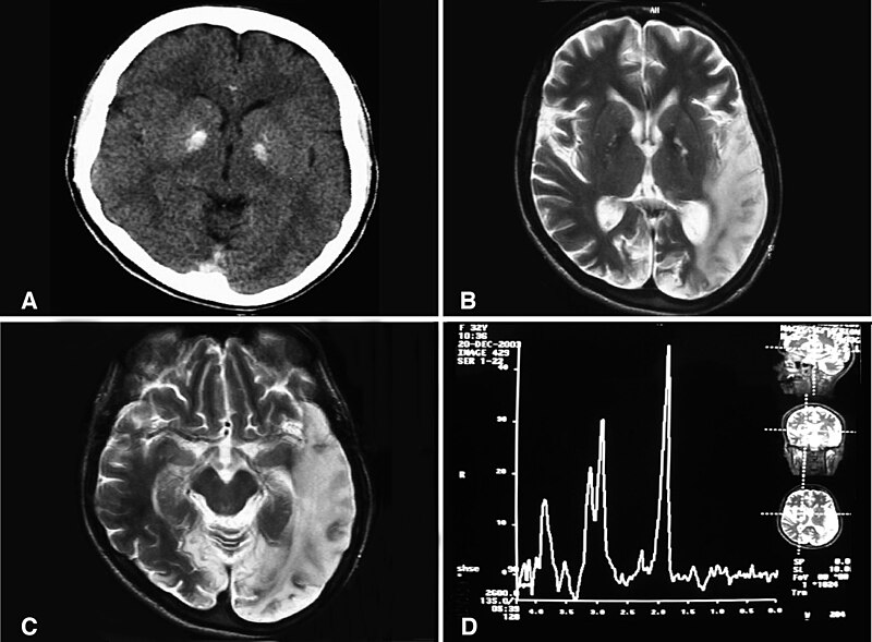
English: (a) A computed tomography brain scan showing bilateral basal ganglia calcification; the cerebellum shows prominent folia indicating mild cerebellar atrophy. (b) Axial T2 brain magnetic resonance image scan showing left temporo-parieto occipital ischemic lesion. (c) Axial T2 brain magnetic resonance image scan showing the extension of the parietal temporal region to the occipital lobe, and also showing a right occipital lesion. (d) Magnetic resonance spectroscopy showing inversion of J-coupling phenomenon at 1.3 ppm, indicating lactate peak. Abu-Amero et al. Journal of Medical Case Reports 2009 3:77 doi:10.1186/1752-1947-3-77 |
| تاریخ | |
| منبع | A patient with typical clinical features of mitochondrial encephalopathy, lactic acidosis and stroke-like episodes (MELAS) but without an obvious genetic cause: a case report |
| پدیدآور | Abu-Amero KK, Al-Dhalaan H, Bohlega S, Hellani A, Taylor RW. |
| اجازهنامه (استفادهٔ مجدد از این پرونده) |
© 2009 Abu-Amero et al; licensee BioMed Central Ltd. This is an Open Access article distributed under the terms of the Creative Commons Attribution License (https://creativecommons.org/licenses/by/2.0), which permits unrestricted use, distribution, and reproduction in any medium, provided the original work is properly cited. |
این پرونده با اجازهنامهٔ کریتیو کامانز Attribution 2.0 عمومی منتشر شده است.
- شما اجازه دارید:
- برای به اشتراک گذاشتن – برای کپی، توزیع و انتقال اثر
- تلفیق کردن – برای انطباق اثر
- تحت شرایط زیر:
- انتساب – شما باید اعتبار مربوطه را به دست آورید، پیوندی به مجوز ارائه دهید و نشان دهید که آیا تغییرات ایجاد شدهاند یا خیر. شما ممکن است این کار را به هر روش منطقی انجام دهید، اما نه به هر شیوهای که پیشنهاد میکند که مجوزدهنده از شما یا استفادهتان حمایت کند.
تاریخچهٔ پرونده
روی تاریخ/زمانها کلیک کنید تا نسخهٔ مربوط به آن هنگام را ببینید.
| تاریخ/زمان | بندانگشتی | ابعاد | کاربر | توضیح | |
|---|---|---|---|---|---|
| کنونی | ۲۸ ژانویهٔ ۲۰۱۰، ساعت ۱۰:۲۰ | | ۱٬۲۰۰ در ۸۸۳ (۱۵۳ کیلوبایت) | CopperKettle | {{Information |Description={{en|1=(a) A computed tomography brain scan showing bilateral basal ganglia calcification; the cerebellum shows prominent folia indicating mild cerebellar atrophy. (b) Axial T2 brain magnetic resonance image scan showing left te |
کاربرد پرونده
صفحهٔ زیر از این تصویر استفاده میکند:
کاربرد سراسری پرونده
ویکیهای دیگر زیر از این پرونده استفاده میکنند:
- کاربرد در ar.wikipedia.org
- کاربرد در de.wikipedia.org
- کاربرد در en.wikipedia.org
- کاربرد در en.wikibooks.org
- کاربرد در es.wikipedia.org
- کاربرد در fr.wikipedia.org
- کاربرد در it.wikipedia.org
- کاربرد در la.wikipedia.org
- کاربرد در ru.wikipedia.org
- کاربرد در sv.wikipedia.org
- کاربرد در tr.wikipedia.org
- کاربرد در www.wikidata.org
- کاربرد در zh.wikipedia.org