
پرونده:Fundus of patient with retinitis pigmentosa, mid stage.jpg
اندازهٔ این پیشنمایش: ۶۹۹ × ۵۹۹ پیکسل. کیفیتهای دیگر: ۲۸۰ × ۲۴۰ پیکسل | ۵۶۰ × ۴۸۰ پیکسل | ۸۷۱ × ۷۴۷ پیکسل.
پروندهٔ اصلی (۸۷۱ × ۷۴۷ پیکسل، اندازهٔ پرونده: ۱۰۷ کیلوبایت، نوع MIME پرونده: image/jpeg)
این پرونده در ویکیانبار موجود است. محتویات صفحهٔ توصیف آن در زیر نمایش داده میشود. |
| توضیح |
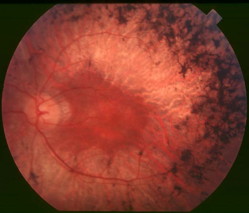
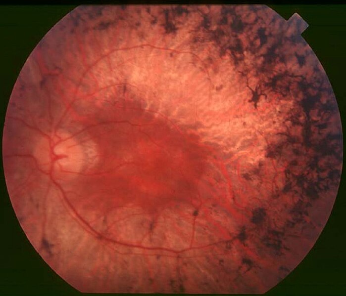
English: Figure 2. Fundus of patient with retinitis pigmentosa, mid stage (Bone spicule-shaped pigment deposits are present in the mid periphery along with retinal atrophy, while the macula is preserved although with a peripheral ring of depigmentation. Retinal vessels are attenuated.) Hamel Orphanet Journal of Rare Diseases 2006 1:40 doi:10.1186/1750-1172-1-40 |
| تاریخ | |
| منبع | Retinitis pigmentosa by Christian Hamel |
| پدیدآور | Christian Hamel |
| اجازهنامه (استفادهٔ مجدد از این پرونده) |
© 2006 Hamel; licensee BioMed Central Ltd. This is an Open Access article distributed under the terms of the Creative Commons Attribution License (https://creativecommons.org/licenses/by/2.0), which permits unrestricted use, distribution, and reproduction in any medium, provided the original work is properly cited. |
این پرونده با اجازهنامهٔ کریتیو کامانز Attribution 2.0 عمومی منتشر شده است.
- شما اجازه دارید:
- برای به اشتراک گذاشتن – برای کپی، توزیع و انتقال اثر
- تلفیق کردن – برای انطباق اثر
- تحت شرایط زیر:
- انتساب – شما باید اعتبار مربوطه را به دست آورید، پیوندی به مجوز ارائه دهید و نشان دهید که آیا تغییرات ایجاد شدهاند یا خیر. شما ممکن است این کار را به هر روش منطقی انجام دهید، اما نه به هر شیوهای که پیشنهاد میکند که مجوزدهنده از شما یا استفادهتان حمایت کند.
تاریخچهٔ پرونده
روی تاریخ/زمانها کلیک کنید تا نسخهٔ مربوط به آن هنگام را ببینید.
| تاریخ/زمان | بندانگشتی | ابعاد | کاربر | توضیح | |
|---|---|---|---|---|---|
| کنونی | ۲ دسامبر ۲۰۱۷، ساعت ۱۰:۱۷ | | ۸۷۱ در ۷۴۷ (۱۰۷ کیلوبایت) | Doc James | Cropped 27 % horizontally and 7 % vertically using CropTool with precise mode. |
| ۲۲ سپتامبر ۲۰۰۹، ساعت ۱۳:۵۲ |  | ۱٬۲۰۰ در ۷۹۹ (۱۲۶ کیلوبایت) | CopperKettle | {{Information |Description={{en|1=Figure 2. Fundus of patient with retinitis pigmentosa, mid stage (Bone spicule-shaped pigment deposits are present in the mid periphery along with retinal atrophy, while the macula is preserved although with a peripheral |
کاربرد پرونده
صفحههای زیر از این تصویر استفاده میکنند:
کاربرد سراسری پرونده
ویکیهای دیگر زیر از این پرونده استفاده میکنند:
- کاربرد در ar.wikipedia.org
- کاربرد در bs.wikipedia.org
- کاربرد در ca.wikipedia.org
- کاربرد در da.wikipedia.org
- کاربرد در en.wikipedia.org
- کاربرد در en.wikiversity.org
- کاربرد در es.wikipedia.org
- کاربرد در eu.wikipedia.org
- کاربرد در fi.wikipedia.org
- کاربرد در fr.wikipedia.org
- کاربرد در he.wikipedia.org
- کاربرد در hy.wikipedia.org
- کاربرد در it.wikipedia.org
- کاربرد در ko.wikipedia.org
- کاربرد در la.wikipedia.org
- کاربرد در or.wikipedia.org
- کاربرد در outreach.wikimedia.org
- کاربرد در pl.wikipedia.org
- کاربرد در pt.wikipedia.org
- کاربرد در ru.wikipedia.org
- کاربرد در sl.wikipedia.org
- کاربرد در sr.wikipedia.org
- کاربرد در sv.wikipedia.org
- کاربرد در th.wikipedia.org
- کاربرد در tr.wikipedia.org
- کاربرد در tt.wikipedia.org
- کاربرد در uk.wikipedia.org
- کاربرد در vi.wikipedia.org
- کاربرد در www.wikidata.org