
پرونده:WW1 fluoroscope operation.jpg
اندازهٔ این پیشنمایش: ۴۲۶ × ۵۹۹ پیکسل. کیفیتهای دیگر: ۱۷۰ × ۲۴۰ پیکسل | ۳۴۱ × ۴۸۰ پیکسل | ۵۴۶ × ۷۶۸ پیکسل | ۷۲۸ × ۱٬۰۲۴ پیکسل | ۱٬۵۶۴ × ۲٬۲۰۰ پیکسل.
پروندهٔ اصلی (۱٬۵۶۴ × ۲٬۲۰۰ پیکسل، اندازهٔ پرونده: ۳۹۶ کیلوبایت، نوع MIME پرونده: image/jpeg)
این پرونده در ویکیانبار موجود است. محتویات صفحهٔ توصیف آن در زیر نمایش داده میشود. |
خلاصه
| توضیح |
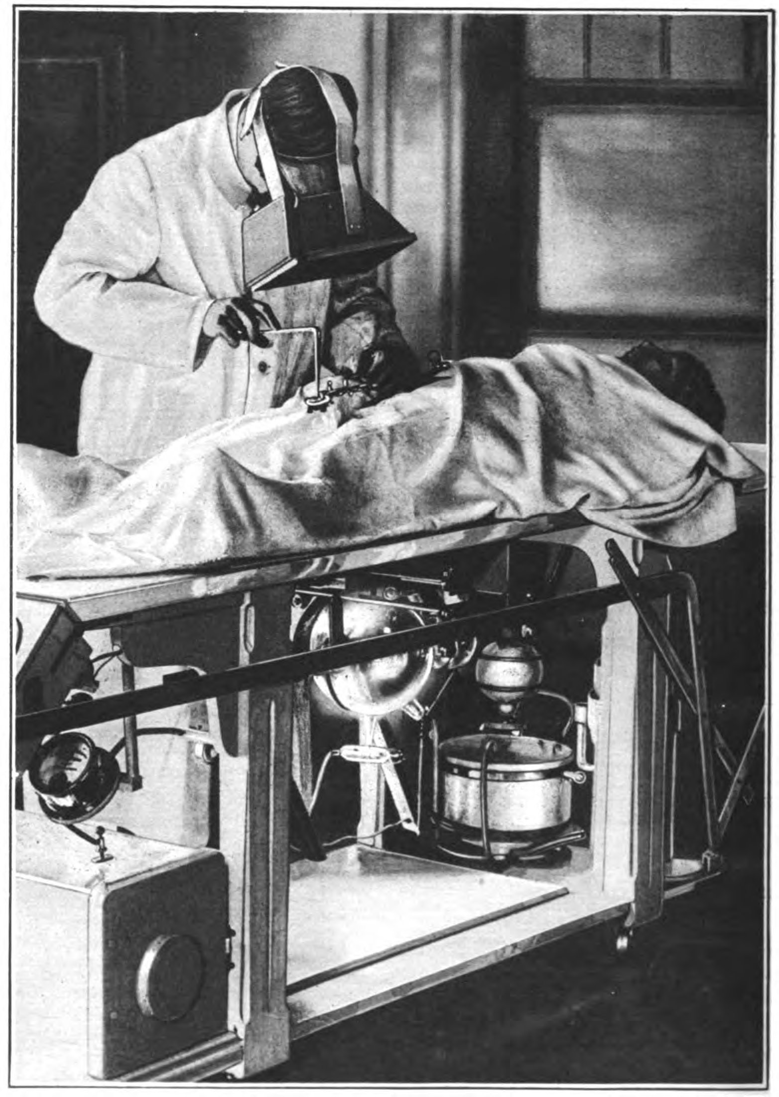
English: Operation on a wounded soldier during World War 1 with the surgeon using a fluoroscope to locate the bullets. An early Crookes x-ray tube visible under the table emits a beam of x-rays vertically through the patient's body. The surgeon wears a large fluoroscope on his face, a screen coated with a fluorescent chemical such as calcium tungstate which glows when x-rays strike it. The x-ray image of the patient's body appears on the screen, with the bullet fragments appearing dark. Although the surgeon is wearing gloves, little protection against radiation appears to be used. X-rays were discovered in 1895, and World War 1 saw the first major use of x-rays in wartime. France and the US sent trucks equipped with early x-ray machines to the front. The photo is credited to Dr. J. P. Hoguet, a surgeon at the Roentgenographic Dept. of the American Ambulance Hospital at Neuilly, France. |
| تاریخ | |
| منبع | Retrieved 12 October 2013 from A. M. Jungmann, "X-rays: Samaritans of war" in Waldemar Kaempffert, Ed., The Book of Modern Marvels, Leslie Judge Co., New York, p. 172 on Google Books |
| پدیدآور | J. P. Hoguet |
اجازهنامه
این پروندهٔ رسانهای در ایالات متحده در مالکیت عمومی قرار دارد. این دربارهٔ آثار ایالات متحده که حقتکثیرشان باطل شده است صدق میکند؛ اغلب به این دلیل که اولین انتشارشان قبل از ۱ ژانویهٔ ۱۹۲۹ روی داده است. برای توضیحات بیشتر این صفحه را ببینید.
|
 | |
 |
ممکن است این نگاره در خارج از ایالات متحده در مالکیت عمومی نباشد؛ این مسئله بهخصوص دربارهٔ کشورها و مناطقی که قانون مدت کوتاهتر را برای آثار ایالات متحدهٔ آمریکا اعمال نمیکنند، همچون کانادا، چین (به جز هنگکنگ و ماکائو)، آلمان، مکیزیک، و سوئیس صدق میکند. آفریننده و سال انتشار اطلاعات ضروری هستند و باید ارائه شوند. برای جزئیات بیشتر، ویکیپدیا:مالکیت عمومی و ویکیپدیا:حق تکثیر را ببینید.
|
تاریخچهٔ پرونده
روی تاریخ/زمانها کلیک کنید تا نسخهٔ مربوط به آن هنگام را ببینید.
| تاریخ/زمان | بندانگشتی | ابعاد | کاربر | توضیح | |
|---|---|---|---|---|---|
| کنونی | ۱۳ اکتبر ۲۰۱۳، ساعت ۱۷:۵۰ | | ۱٬۵۶۴ در ۲٬۲۰۰ (۳۹۶ کیلوبایت) | Chetvorno | User created page with UploadWizard |
کاربرد پرونده
صفحهٔ زیر از این تصویر استفاده میکند:
کاربرد سراسری پرونده
ویکیهای دیگر زیر از این پرونده استفاده میکنند:
- کاربرد در en.wikipedia.org
- کاربرد در ja.wikipedia.org
- کاربرد در uz.wikipedia.org